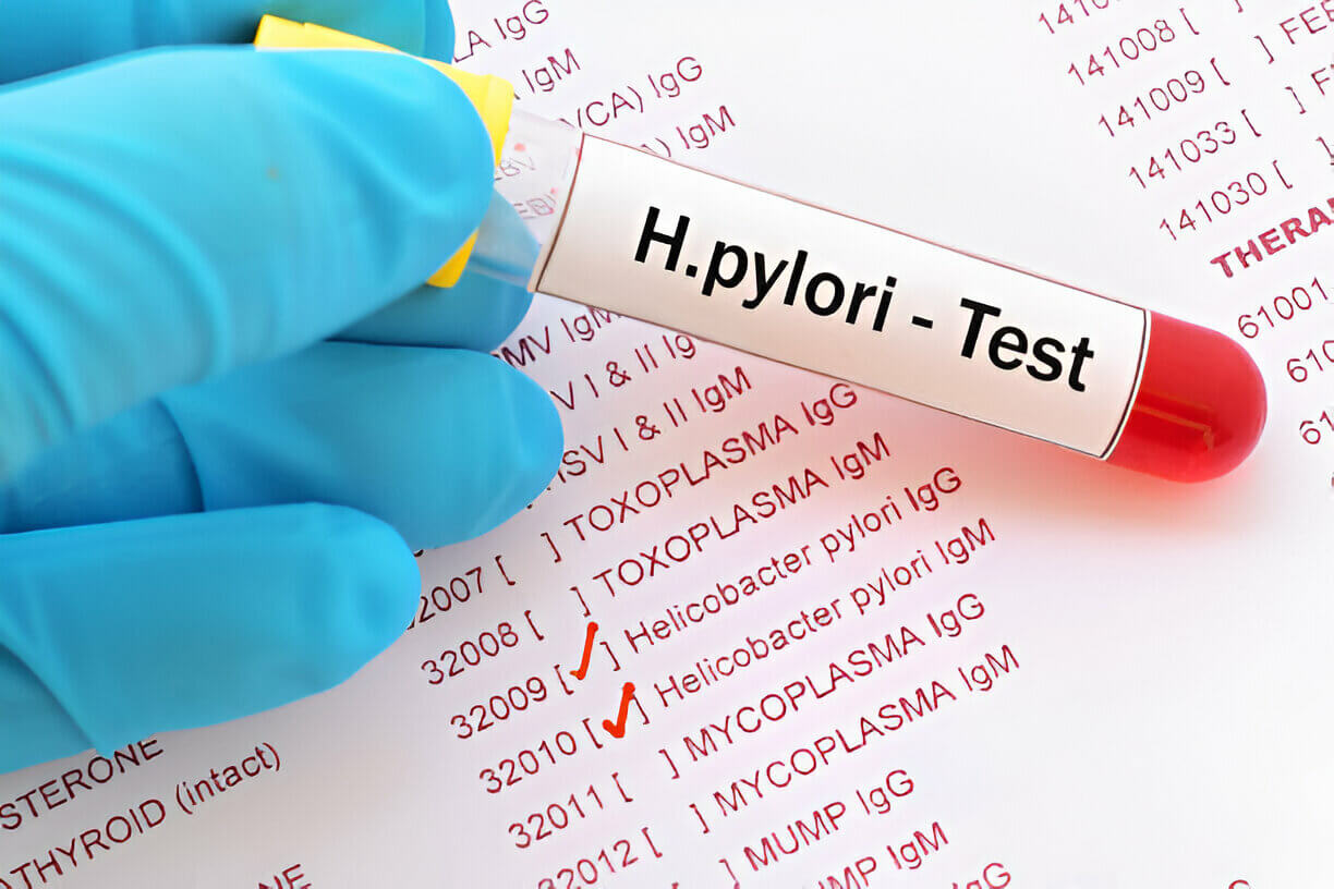
Insights into Helicobacter pylori and Transport Media

In a study conducted by Surg Capt RN Misra, Lt Col M Bhagat, and Dr. N Ahmed from INHS Asvini, Colaba, Mumbai, and CH (CC), Lucknow, the researchers aimed to delineate colonizers from pathogens of Helicobacter pylori (H. pylori) to facilitate appropriate treatment planning. H. pylori is a bacterial species implicated in various gastric conditions, including acute superficial gastritis, chronic atrophic gastritis, gastric carcinoma, and MALT-associated lymphoma. While colonization can occur in normal individuals, treatment is warranted when the organism is associated with virulence factors like vacuolating toxin and cytotoxin, encoded by the vacA and cagA genes, respectively.
What Media Were Used for H. pylori Sample Preservation?
During the study, four gastric biopsies were collected from each patient and preserved in different media for various analyses. The media used included Cary-Blair medium, 10% formalin, Tris-EDTA buffer, and 10% urea broth. Each medium served a specific purpose in the identification and characterization of H. pylori.
What Role Did Each Medium Play?
-
1. Cary-Blair Medium: This medium was used for bacterial culture and isolation of H. pylori. The tissue samples were homogenized in Cary-Blair medium and then inoculated onto Bacto Campylobacter Agar kit Skirrow (Difco) for selective growth of H. pylori.
-
2. 10% Formalin: Tissue samples preserved in 10% formalin were used for histopathological examination. Paraffin sections were stained with hematoxylin-eosin, Giemsa, and toluidine blue to study the tissue morphology, inflammatory cells, and the presence of curved, S-shaped bacteria resembling H. pylori.
-
3. Tris-EDTA Buffer: Samples in Tris-EDTA buffer were utilized for DNA extraction and subsequent PCR analysis. The DNA was extracted using spin column methods, and multiplex PCR was performed to detect the presence of 16SrRNA, vacA, and cagA genes, which are specific markers for H. pylori identification and pathogenicity.
-
4. 10% Urea Broth: Tissue samples were placed in 10% urea broth containing a phenol red pH indicator to perform the rapid urease test. A color change to pink within 30 minutes indicated strong urease activity, suggesting the presence of H. pylori.
What Liquid Was Used for PCR Analysis?
The tissue collected in Tris-EDTA buffer was used for DNA extraction and subsequent PCR analysis. The DNA was extracted using spin column methods with solutions provided in the QIAGEN kit. Multiplex PCR was then performed on the extracted DNA to detect the presence of the 16SrRNA gene, which is specific to H. pylori, as well as the virulence factors vacA and cagA genes.
Why Was PCR Analysis Crucial?
PCR analysis played a crucial role in differentiating commensal H. pylori strains from potentially pathogenic strains. The presence of the vacA and cagA genes, which encode for vacuolating toxin and cytotoxin, respectively, indicates a higher pathogenic potential. By detecting these virulence factors, the researchers could distinguish innocuous bystanders from potentially invasive organisms, aiding in appropriate treatment decisions.
How Did the Transport Media Contribute to the Study?
The use of different transport media allowed for a comprehensive analysis of H. pylori detection and characterization. Cary-Blair medium facilitated bacterial culture and isolation, enabling antibiotic sensitivity testing and further characterization of the isolates. Formalin-fixed samples provided insights into histopathological changes associated with H. pylori infection. The Tris-EDTA buffer preserved DNA integrity for PCR analysis, enabling the detection of specific genes linked to pathogenicity. The urea broth supported the rapid urease test, a widely used screening method for H. pylori.
By employing these various transport media, the researchers could leverage multiple diagnostic approaches, including culture, histopathology, serology, and molecular techniques, to comprehensively assess H. pylori infection and its associated virulence factors. The integration of these methods contributed to a deeper understanding of the disease dynamics and aided in distinguishing colonizers from potential pathogens, ultimately informing treatment strategies.
Mantacc Cary-Blair Agar Gel Transport Swab
By incorporating Manntacc's Cary-Blair Agar Gel Transport Swab into your H. pylori culture protocols, you can enhance the accuracy and reliability of your results while reducing the complexities associated with traditional transport methods. Embrace this cutting-edge solution and unlock new possibilities in the pursuit of better understanding and management of H. pylori infections.

References
Helicobacter pylori in Dyspepsia – Antibiotic Sensitivity and Virulence Patterns


